1Consultant General surgery Armed forces Hospital Southern region Khamees Mushayt, Kingdom of Saudi Arabia
2Former Registrar Armed forces Hospital Southern region Khamees Mushayt, Kingdom of Saudi Arabia
*Corresponding author: Hesham M Haroon, General Surgery Department, Armed Forces Hospital Southern Region, Kingdom of Saudi Arabia.
Citation: Haroon HM, Aljoufi A, Khudhayr E, Shamshad H. (2023) Penetrating Abdominal Injuries in War Victims Challenges and Management Controversies. Genesis J Surg Med. 2(2):1-08.
There is no doubt that penetrating abdominal injuries are big challenge to all surgeons and significant cause of morbidity and mortality worldwide. this problem is more dangerous in war victims due to the complexity of weapons used in the modern battles which have a higher velocity, kinetic energy, and consequently potential to destroy tissues, in addition to toxins and harmful chemicals used. Blast injuries also include most of the mechanisms in trauma.
Penetrating abdominal injuries; Gunshot thoracoabdominal injuries, Complex mesenteric vascular; Major vascular injuries
The most common challenges and controversial issues facing the surgeon when dealing with such cases are:
1. To Operate or Not?
2. How to manage Colonic Injuries.
3. How to manage Complex Hepato-pancreato-duodenal injuries.
4. Management of thoracoabdominal injuries.
5. Management of Mesenteric and Major Vascular injuries.
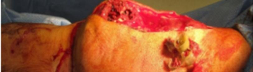
A retrospective review of penetrating abdominal injuries in War victims from August 2015 till August 2019 in Armed Forces Hospital Southern Region in KSA with exclusion of cases with concomitant major chest, central nervous system, and Musculo-skeletal System injuries. A total of 64 patients all are males were seen. Age ranges between 20-35 years old with mean age 28 yrs. Old. All the cases are Gunshot injuries in the form of Bullets (40, 62.5%), or shrapnel’s (24, 37.5%).
All patients have been resuscitated and rehydrated and managed according to the protocol of ATLS (ABCDE), they have been given proper antibiotic cover, antitetanic serum, and properly assessed by CT trauma and all other needed diagnostic modalities.
The most common injured organs are small intestine, Colon, stomach, and liver. Other less common injuries include the spleen, diaphragm, kidney, duodenum, Gall Bladder, Pancreas, and Biliary Tree. From the 64 patients 4 patients (6.25%) has been selected for Non-operative Management one of them has been explored after 24 hours due to generalized abdominal tenderness, tachycardia, and fever we found retroperitoneal left colonic injury and resection done with end colostomy. Two patients had diagnostic laparoscopy (3.125%) which showed liver tears and diaphragmatic injuries and managed laparoscopically. 58 patients (90.6 %) had formal laparotomy exploration where organs most affected were: Small intestine 25 cases (43.1 %), Colon 17 cases (29.3 %), liver 7 cases (12 %), Diaphragm 4 cases (6.8 %), Stomach 3 cases (5.1 %), Duodenum, Pancreas, and CBD 3cases (5.1 %), Major Vascular injuries 2 cases (3.44 %), and Rectum one case (1.7 %).
Mortality was 2 cases (3.125 %) due to major bleeding and Complex Duodena-pancreatic injuries. The overall complication rate was 26.2% with no significant differences between the 4 groups (Table 1), the most common complication was lung related (pneumonia and acute respiratory distress syndrome, 16.38%. An anastomotic leak developed in 2 patients both of them colonic (2.38%).
|
Complication
|
N (%)
|
|
Lung related
|
7(11.47 %)
|
|
Acute Respiratory Distress Syndrome
|
3 (4.91%)
|
|
Anastomotic leak
|
1 (1.64 %)
|
|
Wound related
|
5 (8.19%)
|
|
Total
|
16(26.21%)
|
Table 2: Postoperative complications.
Discussion
Symptoms and signs of penetrating abdominal injuries varies greatly according to the type of weapon used, distance from which injury occurred, which organ affected, location, and number of wounds, however some occult injuries may be overlooked resulting in devastating complications [5,6]. In abdominal war wounds mortality rate dropped from 53 % during world War I to 18-36 % at the end of World War II and went down to 10 % in Vietnam War in some hospital series and 11.5 % in another series [3]. In cases of abdominal wounds was the primary lesion death occurred due to Hemorrhage (60%), Sepsis (25%), and Pulmonary Insufficiency in 15 % [3]. Although Stab wounds are encountered more the overall complication rate was 26.2% with no significant differences between the 4 groups (Table 1), the most common complication was lung related (pneumonia and acute respiratory distress syndrome, 16.38%. frequently in civilian victims than in War victims (3 Times) usually the mortality rate is lower, however approximately 90 % of deaths due to penetrating abdominal injuries are due to gunshot wounds (GSW) [7].
Selective Non-Operative Management (SNOM) in stab wounds has become a popular management modality, provided the patient is hemodynamically stable and there is effective serial physical examination policy in a well-established trauma set-up. In Gun Shot Wounds the SNOM is still controversial issues and depends on several factors: hemodynamic stability of the patient, presence of diffuse abdominal tenderness, associated head and spine injuries, and presence of close monitoring and effective serial physical examination for 12-24 hours. Preferably by the same physician [3]. Good diagnostic modalities should be available like C-T, FAST, and angiography suit to support the decision of Non-Operative management and identifying the patients for Operative management. Gun Shot Wounds of Anterior abdomen, Back, and Buttocks follow the same principles in management [8,9]. In a Study published 2001 by Velmahos et al., 1975 patients with abdominal GSW were included and 1405 of them had anterior GSW, 484 were managed non-operatively (34%). Sixty-five patients received a delayed laparotomy (13.4%) after developing signs and symptoms, but only 48 (9.9%) had significant injuries. Seventeen patients had a non-therapeutic laparotomy (26.2%) [10].
Another prospective study done by Velmahos and his colleagues on 192 patients with GSW to the back over a 12 months period. One hundred and thirty were selected for SNOM four of them received delayed laparotomy but it was non-therapeutic in all of them. SNOM was successful in the remaining 126 (96.9 %). Their clinical examination showed a sensitivity of 100 % and specificity of 95% [5]. Same results were published for GSW to the pelvis and buttocks with rigid sigmoidoscopy was introduced as routine in all cases.
Colon Injuries
Colonic Injuries constitute a big challenge as it has undergone a major change in the last three decades. After World War II all colonic injuries were routinely managed by performing a colostomy until 1970s. This was gradually replaced by primary repair in selected cases then by liberal primary repair in 1990s [7]. In a prospective randomized study of 268 patients they showed that in selected patients primary repair was associated with fewer complications than colostomy (15 %Vs 19 %) (p> 0.05). The Author concluded that specific criteria should be present to have primary closure as the preferred method of treatment:
(1) Preoperative shock never being profound. (2) Blood Loss less than 20 % of estimated blood volume. (3) No more than two intra-abdominal organs injured.
(4) Minimal fecal contamination.
(5) Operation done within 8 hours of injury and wounds of colon and abdominal wall never so destructive to require resection [6].
Demetriades et al., compared primary anastomosis with diversion in a recent prospective multicenter study of 297 patients. 197 of them underwent primary anastomosis compared to 100 in the diversion group. There was no difference in the colon-related complications between the 2 groups. (22 % Vs 27 %, p >0.313) respectively. The Author concluded that in severe colon injuries requiring resection, the method of colon management does not influence the incidence of colon related complications irrespective of presence or absence of any risk factors [11].
Management of Thoracoabdominal Injuries
The right sided thoraco-abdominal injuries with GSW are more likely to be treated with the SNOM provided the patient is hemodynamically stable and no suspicion of visceral or biliary tree injuries in the trauma C-T as the diaphragm can be sealed by the presence of liver on the right side. On the left side the incidence of diaphragmatic injury is higher (59 % Murray et al) Patients with indications for surgery (peritonitis) hemodynamic instability, hemothorax, and cardiac tamponade underwent exploration whereas patients with no indications underwent laparoscopy. Among patients evaluated laparoscopically 10% were found to have diaphragmatic injuries whereas among patients explored 76 % were found to have a diaphragmatic injury [12]. In another Study by the same group the incidence of occult diaphragmatic injuries was 24 %. The Accuracy of the C-T in diagnosing diaphragmatic injuries were 96 % as shown by Maryland Group [12].
Duodenal Injuries
They are uncommon and found in only 3.7 % of all laparotomies for trauma. It merits a special consideration for the following reasons: (I) The retroperitoneal situation of most of the duodenum may make rupture difficult to detect; (II) The duodenum is easily narrowed by sutures; (III) one type of injury, duodenal hematoma requires a different type of management; (IV) It is a lesion that is badly treated. The intraperitoneal rupture produces the syndrome of gut rupture with more pain than usual. The retroperitoneal rupture is more difficult to detect as the symptoms may be diminished in the beginning then after few hours or even one day the patient will start to have severe pain in the epigastrium and back with general appearance of sepsis and high morbidity and mortality. Primary repair or Duodenorrhaphy is successful in most duodenal wounds if discovered early. Duodenal hematoma if discovered during laparotomy should be expressed carefully to avoid mucosal injury as if it happened repair will be difficult, and the patient may be in need for duodenal resection or diversion [13].
However, it may be complex and the management difficult especially when the diagnosis is delayed or when massive injury to the pancreato- Duodeno-biliary complex occurs from penetrating trauma. Late mortality is generally a consequence of infection or multiple organ failure.
Pancreatic Injury
Traumatic injury to the Pancreas is uncommon occurring in 2-3 % of severe abdominal injuries and the prognosis is influenced by the cause and complexity of the pancreatic injury, the amount of blood loss, duration of shock, and speed of resuscitation [14]. Major injuries to the Pancreas are among the most complex challenges a surgeon is likely to encounter. Isolated injury to the Pancreas is infrequent and associated injuries occurs in 50-90 % of cases. Associated injuries include liver lacerations, avulsion of the common bile duct and gastroduodenal, right, and middle colic vessels. Organs commonly associated with pancreatic injuries are: Liver (44 %), stomach (40 %), major vessels (35 %), colon (30 %), thoracic viscera (31 %) [14].
C-T is the imaging investigation of choice, ERCP, and MRCP are helpful diagnostic modalities for diagnosing pancreatic-biliary injuries. Operative priorities are to identify and control the bleeding sources and replacement of losses, and repair of major vascular injuries. Minor contusions or lacerations of the Pancreatic substance does not require definitive management after exclusion of major duct injury which can be suggested by mechanism of injury, lesser sac fluid collection, retroperitoneal bile staining, crepitus or hematoma overlying the pancreas at the base of the transverse mesocolon [14]. Intraoperative evaluation of the pancreas includes assessment of the integrity of the main pancreatic duct, the presence and extent of duodenal injuries, and whether the pancreatic head or duodenum is devitalized, the ampulla is disrupted, or bile duct is injured.
The easiest and most convenient intraoperative radiological test to diagnose biliary or pancreatic duct injury is operative cholangiogram through cystic duct after gall bladder removal or by using butterfly needle to common bile duct. Injury to the distal pancreatic duct is best treated with distal pancreatectomy and splenectomy with over sewing the resected margins [15]. Proximal injury with possible duct disruption is best managed by external drainage provided no devitalization and Ampulla is intact (External Fistula). Combined Pancreatic Head and duodenal injuries are uncommon and usually result from gunshot wounds. Proper intra-operative assessment of the Ampulla, CBD, Pancreatic duct, and duodenum can be achieved by cholangiogram as before. If the CBD and Ampulla are intact, the duodenal laceration is repaired, and the pancreatic injury treated according to the site of injury [13]. Localized ischemia and necrosis at the site of duodenal injury should be debrided before primary closure. In severe duodenal injury with less injury to the pancreatic head so diversion of gastric and biliary contents away from the duodenal repair (Vagotomy + Antrectomy + gastrojejunostomy + CBD exploration and T-Tube. Another alternative is via pyloric exclusion + gastrojejunostomy + T-Tube [14].
Whipple procedure may be suitable in case of severe tissue devitalization with complete disruption of the Ampulla, involving proximal Pancreatic duct and distal CBD or avulsion of the Duodenum and Pancreas. The decision to do Whipple procedure depends on the extent of tissue damage, experience of the surgeon, and hemodynamic stability of the patient [15]. Pancreatic fistula is the most common complication after pancreatic injury and occur in 10-20 % of major injuries. Most fistulas are minor and resolves spontaneously within 1-2 weeks provided adequate drainage has been established. The management of severe proximal pancreatic injuries remain one of the most difficult challenges in abdominal trauma surgery [16]. Major abdominal Vascular injuries need emergency laparotomy to control hemorrhage and reversal of shock. Major vessels involved are vena cava, portal vein, superior and inferior mesenteric vessel, Aorta, and Iliac vessels. Immediate compression and control of the bleeding areas then proper vascular repair [16].
Central or Midline retroperitoneal Hematomas should be explored specially if progressive expansion happened. Pringle maneuver can help to stop bleeding from the liver or peri duodenal areas. Superior mesenteric vessels can be approached from left lateral side at the level of pancreas and every effort should be made to repair or replace the injured part [17]. Etroperitoneal bile staining, crepitus or hematoma overlying the pancreas at the base of the transverse mesocolon. Intraoperative evaluation of the pancreas includes assessment of the integrity of the main pancreatic duct, the presence and extent of duodenal injuries, and whether the pancreatic head or duodenum is devitalized, the ampulla is disrupted, or bile duct is injured.
Conclusion
Penetrating abdominal injuries in the war are more serious than ones in the civilian environment due to brutality of battlefield, and absences of proper medical services, and ideal hospitals. Management of such cases depends on the ATLS protocol, primary survey, and secondary survey in addition to availability of proper resources fixed protocol for the complex injuries and availability of special trained trauma surgeons ready to deal with such conditions.
So, in this study we recommend If the patient is vitally stable and by examination, no signs of surgical abdomen and radiologically no evidence of Organ Injury + Zone I Injury, you can observe the patient under close monitoring with serial examination. If the patient is unstable or there are signs of surgical abdomen, or there is evidence of viscus perforation by CT, you must take the patient immediately for formal exploration laparotomy. If you have stable patient or slightly increase in pulse with equivocal abdominal examination with no evidence of organ injury by CT, you can take the patient for diagnostic laparoscopy.
References
1. Isenhour JL, Marx J. (2007) Advances in abdominal trauma. Emerg Med Clin N Am. 25:713-33.
2. Dickinson ET, Braslow B. (2006) Acute abdominal eviscerations. Cir Esp. 31:70-81.
3. Cardi M, Ibrahim K, Alizai SW, Mohammad H, Garati H, et al. (2019) Injury patterns and causes of death in 953 patients with penetrating abdominal war wounds in a civilian independent non-governmental organization hospital in Lashkargah, Afghanistan, World J Emerg Surg. 21:14:51.
4. Jowan G. Penn-Barwell, Brown KV, Fries CA. (2015) High velocity gunshot injuries to the extremities: management on and off the battlefield: Cur Rev Musculoskeletal Med. 8(3):312–17.
5. Velmahos GC, Demetriades D, Foianini E, Tatevossian R, Cornwell EE 3rd, et al. (1997) A selective approach to the management on gunshot wounds to the back. Am J Surg. 174(3):342-46.
6. Cullinane DC, Jawa Rs, Como JJ, Morris DS, Cheriyan J, et al. (2019) Management of penetrating intraperitoneal colon injuries: A meta-analysis and practice management guideline from the Eastern Association for the Surgery of Trauma. J Trauma Acute Care Surg. 86(3):505-15.
7. Muhammad U Butt, Zacharias N, Velmahos GC. (2009) Penetrating abdominal injuries: management controversies: Scandinavian Journal of trauma. Scand J Trauma Resus. 17:17:19
8. Vertrees A, Greer L, Pickett C, Nelson J, Wakefield M, et al. (2008) Modern management of complex open abdominal wounds of war: A 5-year experience. J Amer Coll Surg. 207(6): 801-09.
9. Oosthuizen GV, Bruce JL, Clarke DL, Kong VY, Odendaal JJ, et.al, (2018) The impact of mechanism on the management and outcome of penetrating colonic trauma, Ann R Coll Surg Engl. 100(2):152-56.
10. Velmahos GC, Salim A, Belzberg H, Vassiliu P, Chan LS, et al. (2001) Selective nonoperative management in 1,856 patients with abdominal gunshot wounds: should routine laparotomy still be the standard of care? Ann Surg. 234(3):395-402.
11. Demetriades D, Murray JA, Nagy KK, Namias N, Wisner DH, et al. (2001) Committee on Multicenter Clinical Trials. American Association for the Surgery of Trauma. Penetrating colon injuries requiring resection: diversion or primary anastomosis? An AAST prospective multicenter study. J Trauma. 50(5):765-75.
12. Murray JA, Demetriades D, Asensio JA, Cornwell EE 3rd. (1998) Vlahos Prospective evaluation of laparoscopy in penetrating injuries to the left lower chest. 187(6):626-30.
13. Kuza CM, Hirji SA, Englum BR, Speicher PJ, Ganapathi Am, et al. (2020) Pancreatic Injuries in Abdominal Trauma in US Adults: Analysis of the National Trauma Data Bank on Management, Outcomes, and Predictors of Mortality. Scand J Surg. 109(3):193-204.
14. R Lochan, G Sen, A M Barrett, J Scott, RM Charnley. (2009) Management strategies in isolated pancreatic trauma. J Hepatobiliary Pancreat Surg. 16(2):189-96.
15. Bozdag Z, Kapan M, Ulger BV, Oguz A, Aldemir M, et al. (2016) Factors affecting morbidity and mortality in pancreatic injuries, Eur J Trauma Emerg Surg. 42(2):231-5.
16. P L van Rooyen, Karusseit VOL, Mokoena T. (2015) Inferior vena cava injuries: a case series and review of the South African experience. Injury. 46(1):71-5.
17. Kamber HM, Neamah HR. (2018) Outcomes of Operative Management of 96 Cases with Traumatic Retroperitoneal Hematoma: A Single-Institution experience. Open Access Maced J Med Sci. 6(11):2128-32.
18. Lund H, Kofoed SC, Hillings JG, Falck-Larcen C, Svendsen LB et al. (2011) High mortality after emergency room laparotomy in hemodynamically unstable trauma patients. Dan Med Bull. 58(5):A4275.
19. da Silva M, Navsaria PH, Edu S, Nickol AJ. (2009) Evisceration following abdominal stab wounds: Analysis of 66 cases. World J Surg. 33:215-19.