Self-Assembling Property of Graphene Derivates Chemico-Physical and Toxicological Implications
Luisetto Mauro1*, Khaled E2, Gamal A Hamid3, Tarro G4, Nili B Ahmadabadi5, Cabianca L6 and Oleg Yurevich Latyshev7
1IMA academy Marijnskaya, Professorship in Toxicology and Pharmacology, Chemical Technology and Chemical Industry Branch Science Branch Italy, Italy
2Professor, Department of Chemistry, Libya Physical Chemistry, University of Benghazi, Libya
3Professor Hematology Oncology, University of Aden, Yemen
4President of the T & L de Beaumont Bonelli Foundation for Cancer Research, Naples Italy
5Nano Drug Delivery, (a Product Development Firm), United States
6Bio-Medical Laboratory Turin Italy Citta’ della Salute, Italy
7IMA academy President, Italy
*Corresponding author: Luisetto Mauro, IMA academy Marijnskaya, Professorship in Toxicology and Pharmacology, Chemical Technology and Chemical Industry Branch Science Branch Italy, Italy.
Citation: Mauro L, Khaled E, Hamid GA, Tarro G, Ahmadabadi NB, et al. (2022) Self-Assembling Property of Graphene Derivates Chemico-Physical and Toxicological Implications. Genesis J Surg Med. 1(2):1-19.
Received: October 6, 2022 | Published: October 21, 2022
Copyright© 2023 genesis pub by Mauro L, et al. CC BY-NC-ND 4.0 DEED. This is an open-access article distributed under the terms of the Creative Commons Attribution-Non Commercial-No Derivatives 4.0 International License. This allows others distribute, remix, tweak, and build upon the work, even commercially, as long as they credit the authors for the original creation.
Abstract
This work start after seeing an recent open letter for transparency related production an quality control technique of mRNA vaccine first signed by Tarro G, Luisetto M and Monsellato ML and an editorial recognized by IMA Marijnskaya academy: graphene and derivates: physico-chemical and toxicology properties in the mRNA vaccine manufacturing strategy Needed specific proof of absence for the regulatory aspects (accepted for publication). Other relevant evidences comes from the work of Giovannini et al related DARKFIED microscope assay of the blood of 1086 symptomatic subjects after vaccination with two types of mRNA vaccine of great interest on this field the work of P Campra and Young RO, Young Me Lee or Ki-Yeob J. Aim of this work is to investigate the self-autoassembly properties of graphene and derivates in order to find relationship in some biotechnological application like mRNA vaccine. After a review part an experimental hypotesys project will be submitted to the researcher to produce a global conclusion. The recent evidences published in last period induced the idea to more deeply study this properties for The clinico-toxicological aspects involved.
Keywords
Self-assempling; Graphene; Graphene GO; Chemico-physicial property; Toxicology clinical effect; Biopharmaceuticals; mRNA vaccine
Introduction
Related various and recent evidence P Campra, Young RO ,Young Me Lee , Ki-Yeob J, Giovannini, et al. and review works Luisetto M, Tarro G it is interesting to observe the self-assembling properties of graphene and its derivates and their implications in clinico-oncological and toxicological field. The characteristic pattern of this innovative material used in many biotechnological applications related To their specific chemico-physical properties are reported in various relevant literature As reported in article “Bio-pharmaceutical manufacturing large scale production process: The graphene-derivates role and m RNA vaccine”. “Used in many bio-medical and other fields like bio-sensors, in water purifing, to remove heavy metals procedure, in diagnostic field but also in extraction, purifying DNA, RNA and other bio molecule, carrier, adiuvant, antibacterial and other biological and industrial use”. In literature it is also possible to see in example.
Materials Today
New Graphene-Based Material Self-Assembles into Vascular Structures, 19 March 2020
“Self-assembly is the process by which multiple components spontaneously organize into larger, well- defined structures. Biological-systems rely on this process to controllably assemble molecular building blocks into complex and functional- materials exhibiting remarkable properties such as the capacity to grow, replicate and perform robust functions. "There is a relevant great interest to develop materials and fabrication processes that emulate those from nature. The ability to build robust functional materials and devices through the self-assembly of molecular components has until now been limited," said team member Yuan hao Wu, who is also at the University of Nottingham - Queen M University London. "This research introduces a new method to integrate proteins with graphene oxide G.O. by self-assembly in a way that can be easily integrated with additive manufacturing to easily fabricate various bio fluidic devices that allow us to replicate key parts of human tissues and organs in the lab".

Figure n1: Color online) Schematic depicting 2 undoped and un-strained freely suspended graphene layers separated by a finite distance (D).

Figure n2: Hexgonal Lattice of Carbons.
Van der Waals Force: a Dominant Factor for Reactivity of Grapheme
Reactivity control of the graphene is an important problem because chemical fictionalization can modulate graphene's unique mechanical, optical, and the electronic properties. Using systematic optical research studies, we demonstrate that van der Waals VDW interaction is the dominant factor for the chemical reactivity of graphene on 2-dimensional (2D) hetero- structures. A significant enhancement in chemical stability of graphene is obtained by replacing the common SiO2 substrate with 2D crystals such as an additional graphene layer, WS2, MoS2, or h-BN. Our theoretical/experimental results show that its origin is a strong van der Waals VDW interaction between graphene layer and the 2D substrate. This results in a high resistive force on the graphene to-ward geometric lattice deformation. We demonstrate that chemical- reactivity of the graphene can be controlled by the relative lattice orientation with respect to the substrates and thus can be used for a wide range of applications including hydrogen storage”.
Self-assembly is a process- mechanims by which a disordered system of pre-existing components forms an organized structure or pattern like a consequence of specific, local interactions among the components themselves, without external direction. When the constitutive components are molecules, the process is named molecular self-assembly. Regarding the self-assembly process in nanoscience it is possible to see.

Figure n3: Conceptual scheme indicating the main stages of the self-assembly process in nanoscience.
Related Graphene Materials
Self-Assembly of organic nano materials and bio materials: the bottom-up approach for functional nanostructures formation and advanced applications “Graphene self-assembly GSA represents a promising and interesting method for micro electronic applications. Recently in last years, graphene micro-patterns (consisting of crossed stripe of single- and 2-layer graphene) have been fabricated by means of the (evaporation-induced) self-assembly technique”
Chinese Chemical Letters
Self-assembly of graphene oxide G.O. nano-sheets in t-butanol/water medium under gamma-ray radiation. “The research works on the properties of graphene oxide (G.O.) in various media has become one of the hottest topics since GO is now the main- principal raw material for graphene-based advanced materials. In this research work, the γ-ray radiation chemistry effect of GO nano-sheets and their self-aggregation behavior in t-butanol/water medium were investigated. The results show that G.O. nano-sheets are reduced and hydroxy-alkylated simultaneously by alcohol free radicals produced by the radiolysis of t-butanol/water solution under γ-ray radiation. The radiation-modified G.O. nano-sheets will self-assemble into a self-standing graphene hydro gel when the pH of solution is lower than 2. A hydroxyl-functionalized free-standing graphene-aerogel is further obtained simply by freeze-drying. This work provides not only a general self-assembly SA mechanism of G.O. nano-sheets in strong acidic alcohol/water media under a high energy radiation, but also a facile and economical preparation method for hydroxy-alkylated graphene-based aerogel.”

Figure n4: Modified grapheme aerogel.
Understanding self-assembly, colloidal behavior and rheological properties of graphene derivatives for high-performance super capacitor Fabrication “Graphene derivatives, like graphene oxide-(G.O.) and reduced graphene oxide (R.G.O.), have been gerat- widely used as promising 2-dimensional nano-scale building blocks due to their interesting properties, cost-effective production, and a good processability. Understanding the intrinsic self-assembling, colloidal, and rheological features of the graphene derivatives is of critical importance to establish the formation- structure-property relationship of graphene-based materials.”
Graphene oxide containing self-assembling peptide hybrid hydrogels as a potential 3D injectable cell delivery platform for intervertebral disc repair applications. “In this reserach study we explore the use of graphene oxide (G.O.) as- like nano-filler for the reinforcement of FEFK.FEFK (β-sheet forming self-assembling peptide) hydrogels. Our results obtained confirm the presence of a strong interactions between FEFK-FEFK and G.O. flakes with the peptide coating and forming short thin fibrils on the surface of the flakes. These strong interactions were found to affect the bulk properties of hybrid hydro gels. At the pH 4 value electrostatic interactions between peptide fibres and the peptide-coated G.O. flakes are thought to govern the final bulk- properties of the hydro-gels while at pH 7, after conditioning with the cell culture -media, electrostatic- interactions are removed leaving the hydro-phobic interactions to govern hydro gel final properties. The GO-F820 hybrid hydro gel, with mechanical properties similar to the NP, was shown to promote an high cell- viability and retained cell metabolic activity in 3D over the 7 days of culture and shown to harbour significant potential as an
injectable-hydrogel scaffold for the in-vivo delivery of NP- cells.”

Figure n5: Chemical structure of FEFKFEFK peptide (A), SEM image of G.O. flakes (Bi) and flake size
distribution (Bii). Bottom: Schematic representation of the formulation route used to prepare peptide/GO hybrid hydrogels (C).
Self-assembly can be classified as either static or dynamic. In static self-assembly, the ordered state forms as a system approaches equilibrium, reducing its free energy. In dynamic self-assembly, patterns of pre- existing components organized by specific local interactions are not commonly described as "self-assembled" by scientists in the associated disciplines. These structures are better described as "self- organized", although these terms are often used inter changeably.
Self-Assembly of Graphene Oxide at Interfaces
“Due to its amphiphilic property, (G.O.) can achieve a variety of nano structures with different morphologies (in ex. membranes, hydrogel, crumpled particles, hollow spheres, sack-cargo particles, Pickering emulsions) by self-assembly. The self-assembly is mostly derived from the self-concentration of G.O. sheets at various interfaces, including liquid-air, liquid-liquid , liquid-solid inter faces.”
“In this research work, a 2-dimensional self-assembled magnetic nano particle-graphene oxide (M.N.P.-GO) nano composite is reported for the detection of D.N.A. Single-stranded D.N.A. (ssD.N.A.) coils, generated through a rolling -circle amplification (RCA) reaction triggered by the hybridization of target oligos and pad- lock probes, have a strong interaction with M.N.P.-G.O nano-tags through several mechanisms including π–π stacking, hydrogen bonding, van der Waals VDW , electrostatic, and hydro phobic interactions. This interaction leads to a hydro dynamic size increase (or aggregation) of MNP-G.O. Nano tags, which can be detected by a simple opto-magnetic setup. Due to the high shape anisotropy, M.N.P.-G.O nano-tags provide stronger opto-magnetic signal than individual MNPs. The avoidance of D.N.A. probes (short ssD.N.A. sequences as the bio-sensing receptor) provides easier material preparation and lower measurement cost.
From real-time measurements of the interactions between MNP-GO and RCA products amplified from a highly conserved Escheric. Coli 16S rD.N.A. sequence, a limit of detection of 2 pM was achieved with a total assay time of 90 min. Even if the non-specific binding force between G.O. and ssDN.A. is much weaker than the specific base-pairing force in a D.N.A. duplex, the proposed method provides a detection limit similar to D.N.A. probe-based magnetic bio-sensors, which can be ascribed to the abundant binding sites between G.O. and ssD.N.A. For target concentrations higher than 100 pM, the MNP-G.O nano tags can be applied for a qualitative naked eye detection strategy based on nanotag-ssD.N.A. flocculation”.

Figure n6: Self-Assembly of Graphene Oxide.
Material and Methods
Whit an observational point of view various relevant literature and figure are reported and analysed. After this review part an experimental project hypotesys will be submitted to researcher in order to produce a global conclusion related the topics of this work. All literature comes from bio medical or other scientific or technological involved database.
“(G.O.) is a unique 2-dimensional (2D) material with interesting physical/chemical properties. GO can be considered as a 2D amphiphilic conjugated polymer, consisting of hydro-philic oxygenated groups and hydro phobic conjugated graphitic- domains. The diverse chemical groups endow GO graphene oxide with high chemical activity to react with other molecules and form new species with graphitic frame work. The amphiphilic properties of G.O. sheets provide them the abilities to self-assemble into 3-dimensional (3D) structure or reduced G.O (rGO) gels with porous micro-structures. The pre -condition of these promising properties of G.O. is its excellent solution-like dispersibility in aqueous or non-aqueous media. These liquid media facilitate the exfoliation of G.O into single-layer sheets and provide the exfoliated G.O sheets with specific chemical environment for functionalization/processing. It is essential to understand the solution- based chemical behavior of G.O graphene oxide, which is important for better application of the G. O. In this review work, we outline the solution-based chemistry of GO mainly in terms of the molecular structure, dispersibility in solvents, solution properties and related processing of GO sheets. This review work aims to systematically present physical/chemical behaviors of G.O in solvents including aqueous and non aqueous solvents, which is helpful for better understanding and application of G.O. graphene oxide materials”[1].
Co-assembly
“We used G.O sheets of 2 different average lateral sizes, including the larger G.O. (GO-L) measuring 10.5 ± 4.5 µm and smaller G.O. (GO-S) of 2.3 ± 0.9 µm, both exhibiting a typical hydro phobic- surface and negatively charged carboxylic- groups on their periphery. We chose ELRs as the protein component because of their modular and disordered nature and the possibility to exhibit different molecular conformations at the different temperatures. The ELK1 sequence is a 51.9 kDa molecule consisting of 24 repeats of single- block made of 4 hydro-phobic penta peptides (VPGIG) and a positively(+) charged (VPGKG) one. This relatively simple molecular design offers an accessible transition temp. (Tt) of 30 °C (at 2% ELK1 in MilliQ water) with clearly different ELR conformations above or below it, as well as medium molecular -weight to enable both cooperative interactions between its charged and hydro-phobic segments as well as with the anionic edge and hydro phobic surface of the G.O . ELRs with similar molecular weight but different levels of charge and hydro-phobicity , as well as a single repeat of an individual block of each of these three ELRs, was used as a controls. Fig reported 1 molecular building blocks (and rationale) for When an ELK1 solution at its Tt (30 °C) is immersed in a larger volume of a G.O. graphene oxide solution, a multilayered membrane of up to 50 µm in thickness develops at the interface around the immersed drop maintaining both solutions separated.
This kind of membrane consists of layers made from both G.O graphene oxide. Sheets and ELK1, with G.O sheets being presented throughout the cross-section of the membrane and ELK1 gradually decreased in concentration from the inside (ELK1- side) to the outside (G.O side) of the membrane. Multi-layered structures are known to emerge from diffusion–reaction mechanisms. We have previously demonstrated that with co-assembling PAs with ELRs, it is possible to trigger a diffusion–reaction mechanism, which generates multi-layered membranes capable of exhibiting dynamic- properties. The same In Similar way, by touching any surface within the first few seconds of formation, the ELK1–G.O. membrane adheres, spontaneously and reproducibly opens, and can be manipulated to grow into tubular structures with spatio-temporal control .In this case, the underlying ELR-G.O mechanism of interaction and supra molecular assembly lead to the growth of a material with remarkably enhanced properties” [2].

Figure n7: A) Table summarizes the key information of the three elastin-like recombinamers (ELRs) used in the study comprising similar molecular-weight but different levels of hydro-phobicity (VPGIG) and positive charge (VPGKG). B) Illustrations of the molecular -structure of a G.O sheet and the supra-molecular organization of ELK1 at its transition temperature (Tt) (30°C) indicating both the charged (red and green) and hydro phobic (brown) segments. C) Schematic of the proposed mechanism of formation illustrating the molecular - supra-molecular conformation of the G.O and ELK1 before and after the co-assembly at the ELK1’s Tt as well as their interaction for membrane formation.
Toxicity studies of six types of carbon nano particles in a chicken-embryo model. “In the present research study, the toxicity of 6 different types of carbon nano particles (CNPs) was investigated using a chicken-embryo model. Fertilized chicken eggs were divided into this following treatment groups: placebo, diamond- NPs, graphite NPs, pristine graphene, small graphene oxide, large graphene oxide GO, and reduced graphene oxide.
Self-assembly of CNPs with albumin amino- acids AA by non-covalent bonds is very efficient, implying that CNPs can be effectively transported into embryos. According to Szmidt et al, lower concentrations (50 and 500 μg/mL) of graphene penetrate the embryo more efficiently than the higher concentrations, due to different NP- dispersion levels. These results were explained by the natural tendency of CNPs to agglomerate when they are coated by albumin- proteins that surround the embryo. In the present RESEARCH study work, we also administrated CNPs to egg -albumin, which gets progressively consumed by the embryo during the development process and is ultimately fully absorbed, ensuring that the whole dose was delivered during embryo-genesis “[3].

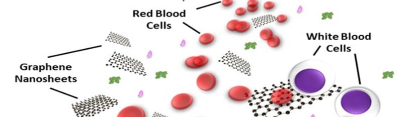
Figure n8: Blood cell aggregation on incubation of graphene with blood from blood compatibility and bio medical applications of graphene.
“In blood, non-covalent adsorption occurs through weak van der Waals VDW forces, hydro phobic, electrostatic, and π–π stacking interactions. The sp2 hybridized honey-comb carbon- lattice of rGO and G.O. is hydro phobic and, interacts with the hydro phobic -regions of proteins, according to the protein geometry. The basal plane of the G.O. is also enriched with π electrons, making π–π stacking interactions possible. At the same time the oxygen -groups of G.O., whose composition is strictly dependent on preparation and storing conditions, allow further hydrogen- bonds and electrostatic bonds. These electrostatic- bonds are strongly influenced by the charge on proteins and by pH-ionic strength of the buffer. Bonding on G.O. can also be mediated by van der Waals VDW-interactions. While the electrostatic- interactions are more pronounced on G.O., both van der Waals WdW and electrostatic -interactions play a major role in the adsorption of proteins on Rg.O due to the increase in the non-functionalized area on the surface. In the following other sections, we will show how fictionalization of the G.O. surface alters protein adsorption and consequently BC -properties”.
Effects of bio-coronated G.O. materials on the blood components BC composition directly influence interactions with the other blood components. The presence of antibodies, complement and clotting factors in the nano-particle BC may activate clotting and coagulation cascades. The BC coating can promote phago-cytosis and elimination from the circulation. We will first consider data on the G.O. interaction with the red blood cells RBC, in Table reported. An intravenously IV injected nano-material is likely to interact first with R.B.Cs rather than other cells, due to their abundance in blood. Hemolysis represents the damage to RBCs that leads to the leakage of hemoglobin into the blood-stream. After hemolysis, the nano-material may adsorb released hemoglobin HB and/or adhere to cell debris, which can increase its likelihood of elimination by macro-phages. Although the literature is contradictory regarding the G.O. effects on R.B.C., when BC is introduced into the framework the results become clearer. Due to the sharp edges of G.O. and Rg.O, hemolytic- effects might be expected in vivo, possibly caused by nano-material blades disrupting cell- membranes, as reported for the G.O. interactions with the bacteria [4].

Figure n9: Main results of G.O interaction with the blood components are summarized in this illustration of the injection of GO graphene oxide flakes in the blood-stream. The formation of the BC (1) prevents the hemolysis of red blood cells RBC (2a). Thrombosis (2b) and interaction with the complement -proteins (2c) are ascribed to GO. In (2d) some of the possible fates after macro-phage encounters are shown: extra- cellular blocking or intra-cellular uptake. The release of cytokines occurs when macro-phages uptake G.O. Aggregates of G.O. in macro-phage cytoplasm induce the production of the pro-inflammatory cytokines. Dendritic-cells fail to present antigens to lymphocytes when they uptake G.O (2e). Lymphocyte activity is not inhibited, and BC protects lymphocytes from apoptosis (2f).
Differential immuno modulatory effect of graphene oxide and vanillin-functionalized graphene oxide nano particles in human acute monocytic leukemia cell line (thp-1). “Graphene and derivatives are emerging as attractive and interesting materials for the bio medical applications: like anti-bacterial, the gene delivery, contrast imaging, and anti cancer therapy applications. It is of fundamental importance to study the cyto-toxicity and the bio compatibility of these materials as well as how they interact with immune-system.
The present research study was conducted to assess the immuno-toxicity of graphene oxide (G.O.) and vanillin-functionalized G.O (V-rGO) on THP-1 cells, a human acute monocytic leukemia cell- line. The synthesized G.O and V-rGO were characterized by using various analytical techniques. Various concentrations of G.O and V-rGO showed toxic effects on THP-1 cells such as the loss of cell viability and proliferation in a dose-dependent manner. Cyto-toxicity was further demonstrated as an increased level of lactate dehydrogenase, loss of mitochondrial membrane -potential (MMP), decreased level of A.T.P. content, and the cell death. Increased levels of reactive- oxygen species ROS and lipid-peroxidation caused redox imbalance in THP-1 cells, leading to increased levels of malon-dialdehyde and decreased levels of anti-oxidants like glutathione, glutathione-peroxidase, super oxide dismutase, and catalase.
Increased generation of ROS and reduced M.M.P. with simultaneous increases in the expression of pro- apoptotic genes and down regulation of anti-apoptotic genes suggest that the mitochondria-mediated pathway is involved in G.O graphene oxide and V-rGO-induced apoptosis. Apoptosis was induced consistently with the significant D.N.A. damage caused by increased levels of 8-oxo-dG and upregulation of various key D.N.A.-regulating genes in THP-1 cells, indicating that G.O. and V-rG.O induce cell death through oxidative stress. As a result of these events, G.O and V-rGO stimulated the secretion of various cytokines and chemokines, indicating that the graphene materials induced potent inflammatory responses to THP-1 cells. The harshness of V-rGO in all assays tested occurred because of better charge transfer, various carbon to oxygen ratios, and chemical compositions in the rG.O. These research findings suggest that it is essential to better understand the parameters governing G.O. and functionalized G.O. in immuno- toxicity and the inflammation. Rational design of safe G.O-based formulations for various applications, including nanomedicine, may result in the development of risk -management methods for people exposed to graphene and graphene family materials, as these nano- particles can be used like delivery agents in various bio medical applications”[6].
“Nano medicines are being developed to treat various diverse diseases; inadvertent or un-intended health effects have to be considered, especially for those targeting cancers. For the cancers, occurrence of metastasis hints an advanced phase of cancer progression, and nano medicines per se should be evaluated for their effects on existing metastatic tumors and triggering the metastases. Graphene-based 2D nano-materials, such as (G.O.), due to its unique characteristics, have been extensively studied for bio medical applications including the cancer therapy. The potential effect of G.O on metastasis has not been determined yet. We found that low-dose G.O could induce significant morphological and structural changes of the cellular membrane within the cancer cells, suggesting an epithelial-mesenchymal transition, with enhanced invasion/migration and the alterations of representative EMT indicators in G.O-treated cells. These changes resulted in enhanced lung -metastasis of cancer cells in various kinds of metastasis models. The mechanistic investigations unveiled that graphene oxide increased the protein levels of the TGF-β receptor, leading to a constitutively activated TGF-β-Smad2/3 signaling path-way that drives the E.M.T. Our findings enhance the understanding of the un-intended side and detrimental effects of G.O. nano-sheets in increasing the progression of metastatic- tumors. So, the likeli-hood of pro-EMT effects upon low-dose GO exposure should be considered when developing G.O. nano medicines [7].
A high dose of G.O. that forms aggregations can block the pulmonary blood -vessels and result in dyspnea and platelet PTL thrombi were observed at high concentrations of 1 and 2 mg/kg body weight via intravenous IV injection [8]. (G.O.) has abundant surfaces oxygen-containing groups like epoxide, hydroxyl, and carboxylic - groups; it can be prepared through the oxidative intercalation and exfoliation of graphite on a mass scale. Owing to the enriched surface functionalities, the G.O is water-soluble and chemically versatile. The surface functional -groups can also provide plenty of reaction sites for linking the nano particles, proteins, enzymes, peptides, bacteria, cells, nucleic acids through the covalent and non-covalent binding.
G.O graphene oxide has been used as a matrix for protein immobilization in different bio technological applications such as fluorescence- or electro chemical-based sensors, labeling and imaging, therapy, and targeted delivery. Non Covalent-interaction (Physical-adsorption), Non-covalent protein adsorption into solid supports represents the most simple and desirable strategy of physical immobilization. The mechanisms of proteins adsorption on G. O graphene oxide is a kind of non- covalent self-assembly including weak Van der Waals VDW forces, hydro phobic, electrostatic, and π-π stacking interaction. These types of attractions between the proteins and graphene oxide G.O. involve solution phase incubation, or direct sonication, followed by a washing step to remove the un-bound proteins. The non-covalent bonds responsible for the interaction between G.O graphene oxide and proteins vary depending on the surface properties of graphene oxide, such as morphology and hydro- phobicity [9].
“Although information on the in vitro and in vivo nano toxicity of graphene nano materials has been increasingly published in the last several years, a complete picture on the bio-compatibility of graphene nano-materials has not been established. The successful applications of graphene nano-materials in nano bio-technology and medicine as well as their effective translation into real clinical utility hinge significantly on a thorough understanding of their nano toxicological profile. Of all aspects of bio compatibility, the hemo-compatibility of graphene nano materials with the different blood constituents in circulatory system is one of the most important elements that need to be well elucidated. Once administered into the biological systems, grapheme nano-materials may inevitably come into contact with the surrounding plasma proteins PP and blood -cells. Crucially, the interactions between these kinds of hematological entities and graphene nano-materials will influence the overall efficacy of their bio medical applications. As such, a comprehensive understanding of hemotoxicity of the graphene nano materials is critically important. he in vitro evaluations of the potential cytotoxic effects of graphene nano-materials have been actively conducted on different human cell -lines, such as human fibroblasts, human umbilical vein endothelial-cells, normal human lung -cells (BEAS-2B), human lung cancer cells (A549), human hepato- carcinoma cells, HeLa cells, and the human breast cancer cells MCF-7. A majority of these investigations have demonstrated the time- and dose-dependent cytotoxicity of graphene nano-materials. Vacuous in vitro experimental and theoric investigations have attributed the cyto-toxicity of both the graphene and its oxygenated derivative G.O. on the mammalian cells and bacteria to cellular membrane penetration, followed by phosphor-lipid molecule extraction from the lipid-bilayer.
G.O. has been demonstrated to possess a high loading capacity for albumin ALB and fibrinogen FIBR in a recent work. While numerous studies have reported observations on graphene nano material-induced a protein conformational change, the under-lying mechanisms are still poorly understood G.O. have a surface area of 25 nm2 and randomly decorated hydroxyl and epoxy-groups on its surface. A carboxyl group was attached to the G.O edges. While having the same surface area, in comparison to G.O graphene oxide, the rGO model possesses fewer oxygenated functional groups. G.O nano-sheets have been reported to possess a strong thrombus-inducing potential and considerable thrombo-genecity.
They could trigger the activation of platelets PTL and their strong aggregatory response similar to that evoked by thrombin, an active physiological platelet agonist. The platelet activation by G.O .was suggested to be extensively dependent on the surface charge distribution of G.O. graphene oxide as it was revealed that, in contrast to G.O, rGO with reduced surface charge density was less capable in activating and aggregating platelets PTL. The pro-thrombotic characteristic of GO nano-sheets was further verified through the occurrence of significant pulmonary thrombo-embolism after their intravenous IV administration in mice [21].

Figure n10: Nano-bio interactions of graphene nanomaterials with various blood plasma proteins and cells.
This critical review work aims at giving insights in the spontaneous tendency of the proteins and their constitutive parts to adsorb on graphitic nano-materials (GNM s) through non-covalent interactions occurring in their interfaces [22].
This work collects studies on the toxic effects of GFNs in various organs and cell models. We also point out that various factors determine the toxicity of GFNs including lateral size, surface structure, functionalization, charge, the impurities, aggregations, corona effect. Various typical mechanisms underlying GFN toxicity have been revealed, for instance, physical destruction, oxidative stress, D.N.A. damage, inflammatory response, apoptosis, autophagy, and the necrosis. In these kind of mechanisms, (toll-like receptors) TLR-, TGF-β-and TNF-α dependent-pathways are involved in the signaling pathway network, and oxidative stress plays a crucial role in these kind of path ways [22].
Toxicity of graphene family nano-particles the dose, shape, surface- chemistry, exposure route, and purity play important roles in differential toxicity of GFNs. Different various authors have used various toxicity tests to evaluate the toxicity of GFNs. Studies have been conducted to find out the toxicity of GFNs on different cellular/animal- models, including stem cells, HeLa cells, HepG2 cells, bacteria, Drosophila melanogaster, Zebra-fish, marine organisms, rats, mice, and mammalian cells. Cytotoxicity tests indicated that the Rg.O can damage cells with direct contact. In this part of the paper, an attempt has been made to compile the recent and up-to-date research studies related to toxicological aspects of GFNs to different models [23].
“The peculiar features of these cases were the availability of macroscopic and micro-scopic autopsy findings. The main macro-scopic finding was that venous- thrombosis was much more wides-pread and catastrophic than diagnosed by imaging during the life. Microscopic findings showed vascular thrombotic occlusions occurring in the micro-circulation of multiple organs and increased inflammatory-infiltrates [24]. Post mortem investigations of fatalities after COVID-19 vaccination are particularly relevant with regard to the detection of anaphylaxis, VITT, and myocarditis.
Vaccine-Induced Immune Thrombotic Thrombo-Cytopenia (VITT)
VITT is characterized by thrombo-cytopenia, combined with thrombosis in most cases. Thrombosis can occur in the both the arterial and, more common, venous system. A distinctive feature of VITT is thrombosis in un-usual locations. These include CVT, as well as splanchnic- venous thrombosis [25].
“In this research work, for the first time, we studied the in vitro and in vivo interactions of a relatively new derivative of graphene, graphene-nanopores (G.N.Ps) in the mammalian systems, to systematically elucidate the possible mechanism of their toxicity over time. Heart tissue showed chemo-dectoma, toxic myocarditis, reddish brown atrophy; yellowish- brown pigments suggesting lipofuscin-granules as remnants of the cell organelles and cytoplasmic-material” (26).

Figure n11: Toxic effects of graphene nanopores on lung cancer cells and biological tissues.

Figure n12: The effect of G.O, rGO, and rGO-PEG on the platelet activation: Graphene nano derivatives were co-incubated with platelet PTL rich plasma at 50 µg/mL. Platelet activation was induced by the addition of 2 µmol/mL of adenosine diphosphate (ADP). The Platelet PTL aggregates are pointed by the blue arrows. One representative picture reflects the results of 3 independent experiments. G.O; rGO reduced graphene oxide; rGO-PEG, PEGylated reduced graphene oxide; ADP, adenosine-diphosphate.

Figure n13: Carbon Nano particles.

Figure n14: Scanning electron microscopy image shows platelet PTL activation by multi wallled CNTs (M60). Platelets were incubated with 100 µg/mL of M60 at 37°C for 15 minutes under gentle agitation. The images are representative of at least three individual experiments with platelets PTL from different donors.
Experimental Project Hypothesis
Results and Discussion
Conclusion
Conflict of Interest
Ethical Consideration
References
Genesis Scientific Publication is licensed under CC BY-NC-ND 4.0![]()
![]()
![]()
![]()
